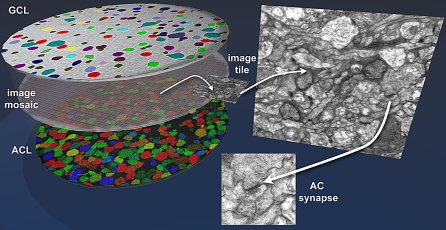
Figure 15. The Retinal CN Mapping Framework.
Canonical fields of rabbit retina are being sectioned from the GC to the AC layers at 70 nm and tiled mosaics acquired for volume assembly. Bounding the ssTEM set are classified sets of GCs (top) and ACs (bottom) whose processes enter the field and can be tagged and tracked. The GC patch is shown as a theme map and the AC patch as a γ.AGB.E :: rgb mapped image. At 5,000× it is possible to unambiguously identify both conventional and ribbon synapses as well as most gap junctions that exceed ≈200 nm in lateral extent. As CMP can be performed on sections as thin as 40 nm, selected molecular signals can be intercalated into ssTEM sets without significant disruption of volume builds by saving spaced sections for CMP, using them if needed, or reinserting them as ssTEM elements if not.

