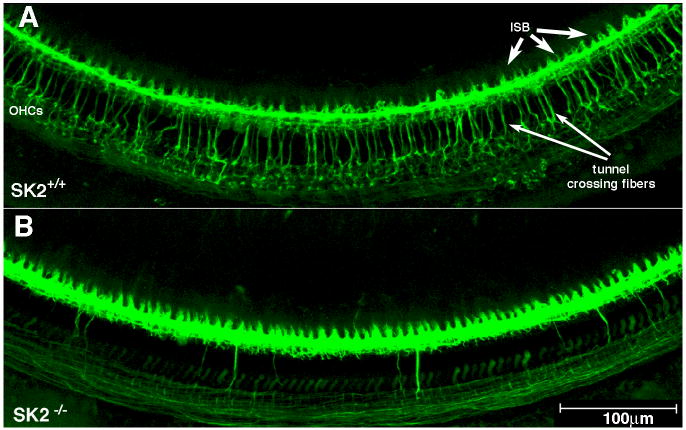
Fig. 2. Olivocochlear fibers degenerate in SK2-/- mice.
Immunostaining for neurotubules and synaptic vesicles (anti-TuJ and anti-synaptophysin; same color) reveals the efferent innervation in a wild type mouse (A) and the loss of efferent innervation in an SK2-/- mouse (B). A: Wild type mice show numerous olivocochlear fibers crossing the tunnel of Corti to the outer hair cells (OHCs; long arrows). The inner spiral bundle (ISB, short arrows), which carries olivocochlear fibers to the inner hair cell area, is also labeled. B: In SK2-/- mice, the number of tunnel crossing fibers and olivocochlear terminals is greatly reduced. Scale bar applies to both panels.

