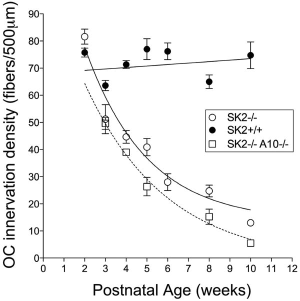
Fig. 4. Quantification of olivocochlear degeneration over postnatal time.
Counting olivocochlear tunnel-crossing fibers reveals a progressive degeneration in SK2-/- (open circles) and SK2-/-/α10-/- (open squares) compared to wild type (filled circle). SK2-/- mice begin with approximately the same number of fibers as wild type mice (2 weeks of age), but steadily decline in numbers over 10 weeks. The double SK2-/-/α10-/- mice exhibit a similar degeneration profile, but the rate of degeneration is increased. Means and SEMs are shown. Data are from the middle of the cochlea in 4-7 cochleae of each genotype at each age.

