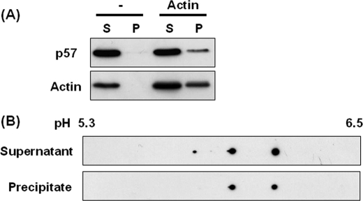
FIGURE 7.
Co-sedimentation of p57/coronin-1 with F-actin. PMA-treated HL60 cells were homogenized in G-actin buffer and centrifuged at 200,000 × g for 60 min to remove insoluble materials. The supernatant was mixed with or without G-actin (5 μg) and then with actin polymerizing solution, and the mixture was incubated at 25 °C for 120 min. After centrifugation at 200,000 × g for 60 min, the supernatant and precipitate were analyzed by one- and two-dimensional gel electrophoresis and immunoblotting. A, the supernatant (S) and precipitate (P) were analyzed by SDS-PAGE/immunoblotting using anti-p57/coronin-1 and anti-actin antibodies. B, the supernatant and precipitate were analyzed by two-dimensional gel electrophoresis/immunoblotting using anti-p57/coronin-1 antibody.

