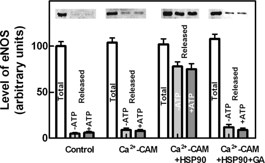
FIGURE 3.
Mobilization of eNOS from isolated endothelial cell membranes. Isolated bEnd5 endothelial cell membranes prepared as described under “Experimental Procedures” were incubated for 30 min at 4 °C in HEPES buffer alone (Control) or containing 0.1 mm CaCl2 + 1 μm CAM (Ca2+-CAM) or containing 0.1 mm CaCl2 + 1 μm CAM + 15 μg of purified HSP90 (17) in the absence (Ca2+-CAM + HSP90) or in the presence (Ca2+-CAM + HSP90 + GA) of 100 nm geldanamycin, and centrifuged at 100,000 × g for 15 min. The supernatants (Released) and the membranes (Total) of each sample were submitted to SDS-PAGE and Western blot analysis and eNOS was detected using the specific antibody. These experiments were performed in the absence (-ATP) or in the presence (+ATP) of 2 mm Mg-ATP, together with 10 mm phosphoenolpyruvate and 2 μg of purified pyruvate kinase, to maintain the level of ATP. The immunoreactive bands were quantified as described under “Experimental Procedures.” The values of each quantification are the arithmetical mean ± S.D. of three different experiments.

