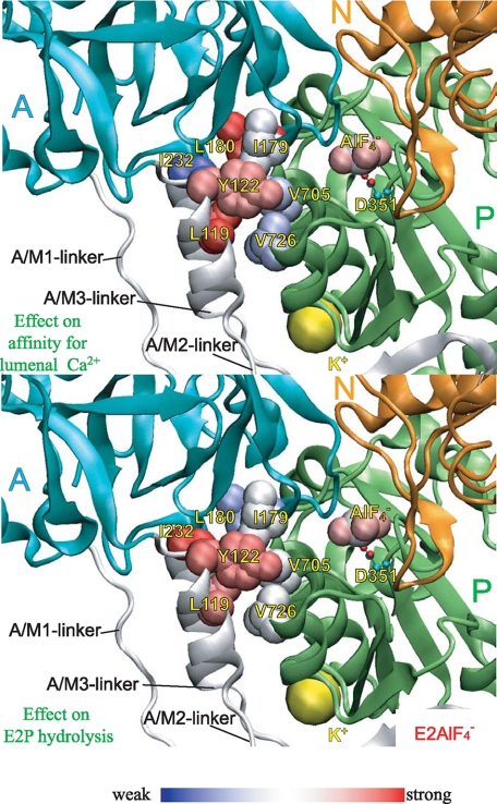
FIGURE 10.
Strength of the mutational effects of seven residues in
Tyr122-hydrophobic cluster on E2P hydrolysis and lumenal
Ca2+ affinity. The detailed structure at Y122-HC is shown with
 (the analog for
E2∼P, the transition state of the E2P hydrolysis
(21), PDB code: 1XP5
(15)). The seven residues
involved in Y122-HC (Tyr122/Leu119,
Ile179/Leu180, Ile232, and
Val705/Val726),
 bound at the phosphorylation site
Asp351, and the bound potassium ion are shown by van der Waals
spheres. The seven residues in Y122-HC are colored
differently based on the strength of the retardation of the E2P
hydrolysis rate (lower panel) and that of the increase in the lumenal
Ca2+ affinity (upper panel). The color changes
gradually from red for the strongest effects to blue for
weakening.

