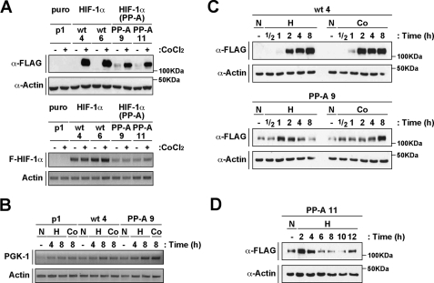
FIGURE 2.
HIF-1α(P402A/P563A) is transiently stabilized in response to hypoxia. A, HIF-1α double proline mutant expressed in stable cell lines is degraded at normoxia. A, upper panel, analysis of mHIF-1α protein levels in stable cell lines expressing wild-type (wt 4 and 6) or HIF-1α(P402A/P563A) (PP-A 9 and 11). Whole cell extracts were separated by SDS-PAGE and analyzed by immunoblotting using anti-FLAG (α-FLAG), or anti-actin (α-Actin) antibodies. Cells were treated with CoCl2 as indicated (+). A pEFIRESpuro-transfected cell line is also shown (p1). A, lower panel, RNA levels of mHIF-1α wild-type or double proline mutant analyzed by semiquantitative RT-PCR. Levels of transcripts were evaluated using primers to FLAG-HIF-1α, and actin. B, RNA levels of PGK-1 are up-regulated in cells expressing HIF-1α or HIF-1α(P402A/P563A). Levels of transcripts were evaluated by semiquantitative RT-PCT using primers to PGK-1, and actin. C, a transient stabilization of HIF-1α(P402A/P563A) is observed at early time points of hypoxia treatment. Stable cell lines (wt 4 and PP-A 9) were treated with CoCl2 (Co) or hypoxia (H) during the indicated times or kept at normoxia (N). Whole cell extracts were separated by SDS-PAGE and analyzed by immunoblotting using anti-FLAG (α-FLAG), or anti-actin (α-Actin) antibodies. D, a different stable cell line expressing HIF-1α double proline mutant confirms the transient regulation of the protein at hypoxia. The stably transfected cell line PP-A 11 was treated with hypoxia (H) as indicated, and HIF-1α expression was analyzed as in C.

