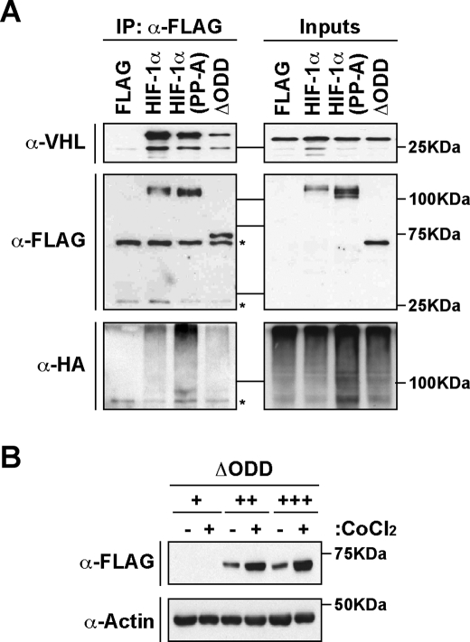
FIGURE 7.
Binding of pVHL to HIF-1α occurs independently of Pro402 and Pro563. A, pVHL binds and promotes ubiquitination of HIF independently of the ODD domain. HEK 293 cells were transfected with pFLAG, pFLAG-mHIF-1α, pFLAG-mHIF-1α(P402A/P563A), or pFLAG-mHIF-1α(391Δ628) (ΔODD), together with pVHL and pHA-ubiquitin. Whole cell extracts (Inputs) and immunoprecipitated FLAG-tagged proteins (IP) were prepared and analyzed by immunoblotting using anti-FLAG (α-FLAG), anti-hemagglutinin (α-HA), or anti-VHL (α-VHL) antibodies. (Denoted unspecific bands are marked with asterisks.) B, pVHL binding correlates with degradation of HIF-1α apart from the ODD domain. HEK 293 cells were transfected with 200 ng (+), 400 ng (++), and 600 ng (+++) of plasmids encoding pFLAG-mHIF-1α(392Δ622) (ΔODD) and exposed to normoxia (-) or CoCl2 (+) for 12 h. Whole cell extracts were separated by SDS-PAGE and analyzed by immunoblotting with anti-FLAG (α-FLAG), or anti-actin (α-actin) antibodies.

