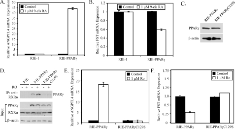
FIGURE 4.
PPARγ requires dimerization with RXRα for repression of follistatin expression. A and B, RIE-1 and RIE-PPARγ cells were treated with 1 μm 9-cis-retinoic acid 9 (9-cis RA) for 6 h. qPCR was used to measure follistatin (A) and ANGPTL4 (B) mRNA expression. C, Western blot of 20 μg of total cell lysate prepared from RIE-1 cells stably expressing WT PPARγ or PPARγC129S. D, nuclear extracts (500 μg) from RIE-1, RIE-PPARγ, and PPARγC129S cells treated with or without rosiglitazone for 6 h were immunoprecipitated with an anti-RXRα antibody and stained for expression of PPARγ by Western blot (lower panel). The Western blot in the lower panel represents 20 μg of the input nuclear extracts stained for PPARγ, RXRα, and β-actin. E and F, abundance of follistatin (E) and ANGPTL4 (F) was measured in cultures of RIE-PPARγ and RIE-PPARγC129S cells treated with vehicle or rosiglitazone (1 μm) for 6 h. Abundance of mRNA was normalized to GAPDH and expressed as -fold change relative to the control. Each bar represents mean ± S.D., n = 3.

