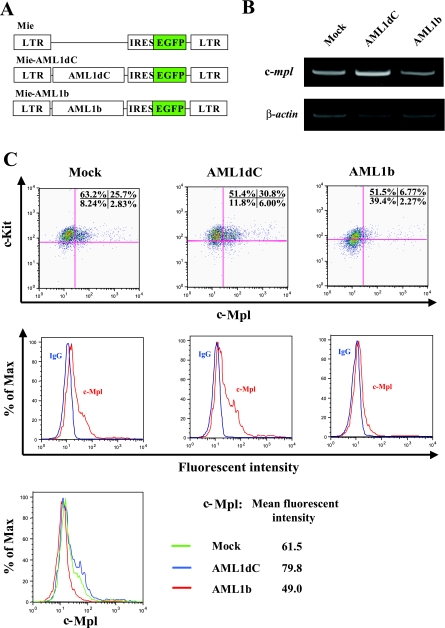
FIGURE 4.
Analysis of the c-Mpl expression in murine hematopoietic stem/progenitor cells. A, structure of Mie (Mock), Mie-AML1dC, and Mie-AML1b retroviruses. B, 2 days after retroviral transfection, GFP+ cells were sorted and subjected to RT-PCR to examine the expression of c-mpl and β-actin mRNA. C, at the same point, the surface phenotype of GFP+ fraction of Mie, Mie-AML1dC, and Mie-AML1b-transduced cells was examined by FACS. Dot plots of cell-surface expressions of c-Kit and c-Mpl (upper panels) are shown. Histogram plots and mean fluorescent intensities of c-Mpl expression (middle and lower panels) are shown. LTR, long terminal repeat; IRES, internal ribosome entry site.

