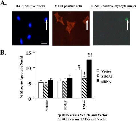
FIGURE 7.
S100A6 modulation of TNF-α-induced myocyte apoptosis as assessed by TUNEL staining. A, representative photomicrographs (×400) of 4′,6-diamidino-2-phenylindole (DAPI)-nuclei (left) (bar = 10 μm), MF20-stained cardiac myocytes (middle), and TUNEL-positive (right) cardiac myocyte(s) treated with TNF-α (5 ng/ml). White arrow indicates myocyte apoptotic nucleus as visualized by TUNEL staining. B, myocyte cultures were transfected with either an S100A6 expression plasmid or an siRNA duplex against S100A6 and treated for 48 h with either PDGF (5 ng/ml), TNF-α (5 ng/ml), or vehicle. TUNEL-positive myocyte nuclei were counted and expressed as percentage of total myocyte nuclei. The results are the mean ± S.E. from random fields in blinded experiments. A minimum of 10 high power fields were scored per experiment of six different experiments. *, p < 0.05 versus vehicle/vector.!, p < 0.05 versus TNF-α/vector.

