FIGURE 1.
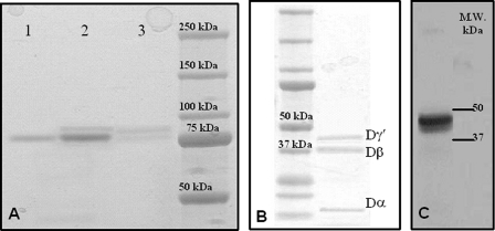
SDS-PAGE of purified fibrinogen fragment D. A, gel was 4–12% polyacrylamide under nonreducing conditions. Lane 1, purified fibrinogen fragment D; lane 2, total fibrinogen fraction eluted by 50 mm sodium phosphate, 80 mm Tris, pH 6.80, in the first chromatographic step using DEAE-Sepharose (see text); lane 3, fraction eluted by 0.5 m NaCl in 20 mm Tris-HCl, pH 8.0, in the second chromatographic step using DEAE-Sepharose (see text). The molecular mass markers are indicated on the right. B, gel was 4–12% polyacrylamide under reducing conditions. The sample was fragment D* obtained from the DEAE chromatography. The component with a molecular mass of ≈41,000 kDa is the elongatedγ chain fragment contained in fragment D*. The other bands pertain to theβ chain region of fragment D* (37.6 kDa) and theα chain fragment (12 kDa). The faint band below the Dγ* may be a minor fragment produced by plasmin digestion, possibly generated by cleavage at Ser86 of the γ chain (61). The molecular mass markers are indicated on the left. C, Western blot of the fragment D* sample shown on the left. Detection of the γ′ chain was obtained using the mouse monoclonal antibody 2.G2.H9, raised against the peptide sequence VRPEHPAETEYDSLYPEDDL of human fibrinogen elongated γ′ chain.

