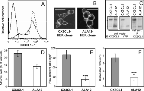
FIGURE 7.
Adhesive properties of HEK clone cells expressing CX3CL1-YFP or ALA12-YFP. A, parental HEK cells (continuous line), stable HEK clone expressing CX3CL1-YFP (fine dotted line), and ALA 12-YFP (heavier dotted line) were stained with phycoerythrin (PE)-labeled murine anti-CX3CL1 mAb and analyzed by flow cytometry. B, the two clones were observed with a Leica SP2-AOBS confocal microscope (bar = 50 μm) C, the two clones were analyzed by SDS-PAGE and Western blot (IB) using antibodies against CX3CL1 (left) or YFP (middle). For immunoprecipitation of antigen located at the cell surface (right), 2 × 10⁶ cells of both clones were incubated in suspension for 1 h at 4 °C with 5 μg of murine mAb anti-human CX3CL1 in PBS plus 0.2% bovine serum albumin. After two washings in cold PBS, the cells were lysed in lysis buffer (150 mm NaCl, 50 mm Tris-HCl, pH 8, 1% Nonidet P-40 (Sigma), Complete protease inhibitor mixture (Roche Diagnostics). The immune complex was separated on protein-G-agarose (Sigma) and analyzed by Western blotting, with a goat anti-human CX3CL1. D, HEK-CX3CL1 or HEK-ALA12 clone cells (105 cells/well) labeled with carboxyfluorescein succinimidyl ester (1 μm, 30 min, 37 °C) were deposited onto confluent CX3CR1-CHO cell or parental CHO cell monolayers. At the end of incubation period and after washings, the plate was read at 535 nm as described under “Experimental Procedures.” The adherent cells are expressed as percentage of total cells minus the mean background corresponding to the number of HEK-CX3CL1 or ALA12 cells adhering on parental CHO cells. E, HEK clone cells expressing CX3CR1 were suspended and assayed for adhesion in a parallel plate laminar flow chamber with coverslips coated with adherent HEK-CX3CL1 or HEK-ALA12 clones as described under “Experimental Procedures.” Adhering cells were counted after 10 min over four fields (mean ± S.E.). The nonspecifically adhering cell numbers were obtained in the same conditions after the addition of 100 nm CX3CL1-CD in HEK-CX3CR1 cells before injection into the flow chamber. The difference between the number of cells specifically adhering either on CX3CL1 or on ALA12 was significant (***, p = 0.002). This result was characteristic of three independent experiments. F, HEK-CX3CL1 or HEK-ALA12 clones were assayed for adhesion by the dual pipette aspiration technique with HEK-CX3CR1 clone cells as described under “Experimental Procedures.” The dissociation force was evaluated after 4 min of adhesion (mean ± S.E., n = 14). The difference between adhesion of the CX3CL1/CX3CR1 and ALA12/CX3CR1 pairs was significant (***, p = 0.008). nN, nanonewton.

