FIGURE 3.
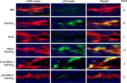
15d-PGJ2 induces p53 protein expression in vascular ECs. Mouse corneas were subjected to alkali trauma and at 3 days later treated with either mock transfection reagent (virus-derived amphipathic peptide alone), control siRNA, or p53 siRNA transfection mixture. After recovery for a further 24 h, the corneas were treated with PBS or 20 μm 15d-PGJ2 for a further 8 h. The corneas were then harvested and subjected to immunofluorescence for eNOS (red; specific for vascular endothelium) and p53 (green). Nuclei were stained by Hoechst 33342 (blue). p53 localized in the nucleus (pale blue) and co-localized with the eNOS (yellow) is shown in merged images. Representative data from experiments are shown. Magnification, ×40. Bar, 75 μm.

