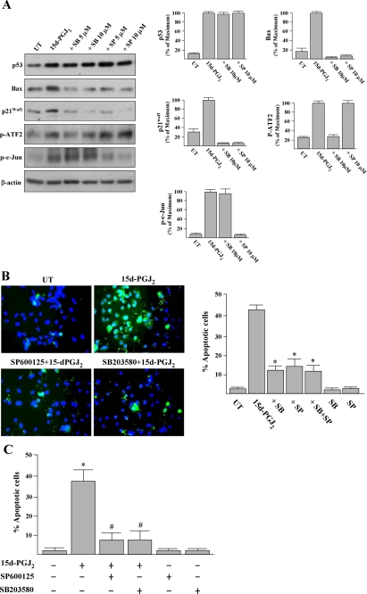
FIGURE 8.
Inhibitors of p38 MAPK and JNK prevent 15d-PGJ2-induced apoptosis and the induction of Bax and p21Waf1. A, HUVECs were pretreated with SB203580 (SB; p38 MAPK inhibitor) or SP600125 (SP; JNK inhibitor) for 1 h and then treated with 15d-PGJ2 for 10 h. Cells were harvested and subjected to Western blot analysis with antibodies as indicated. Representative blots and densitometric analysis from three independent experiments are shown. B, untreated (UT)- and 15d-PGJ2-treated cells were also analyzed using the annexin V-FITC apoptosis detection kit according to the in situ staining protocol after treatment for 16 h. A representative result of four independent experiments is shown. Double-staining with an annexin V-FITC and the fluorescent dye Hoechst 33342 was employed to visualize the apoptotic cells (green) and nuclei (blue), respectively. The percentages of apoptotic cells were quantified. *, p < 0.001 versus 15d-PGJ2-treated cells. C, inhibitors of JNK or p38 MAPK attenuate 15d-PGJ2-induced vascular EC apoptosis. Animal treatment is described under “Experimental Procedures.” After treatment for 24 h, corneas were harvested, and apoptotic endothelial cells (nuclei surrounding annexin V) were quantified as description of Fig. 4. *, p < 0.05 versus PBS-treated corneas. #, p < 0.05 versus 15d-PGJ2-treated corneas.

