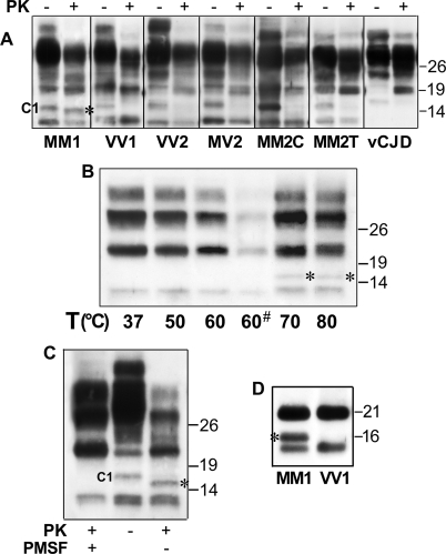
FIGURE 5.
WB analysis of PrPSc digestion in different partially denaturing conditions. A, immunoblot analysis of PrPSc from frontal cortex of all sCJD subtypes and vCJD. Samples were incubated in GdnHCl 1 m for 1 h and untreated or treated with PK. B, immunoblot analysis of PrPSc from frontal cortex of an MM1 case digested with PK at different temperatures. The incubation time was 30 min for all samples but one (marked with an asterisk), which was incubated for 1 h. C, immunoblot analysis of PrPSc from frontal cortex of an MM1 case. Standard (i.e. “PMSF+”) and “PMSF–” conditions, as indicated under “Experimental Procedures,” are compared. Membranes were probed by the 2301 antiserum. D, immunoblot analysis of PrPSc from frontal cortex of an MM1 and a VV1 subject. All samples are in PMSF–conditions and deglycosylated. Membranes were probed by the 2301 antiserum. The 16-kDa fragment generated in denaturing conditions (DCF 16) is marked with an asterisk. All results were reproduced twice with samples from nine sCJDMM1/MV1 and at least three subjects for each of the other groups. Approximate molecular masses are in kilodaltons.

