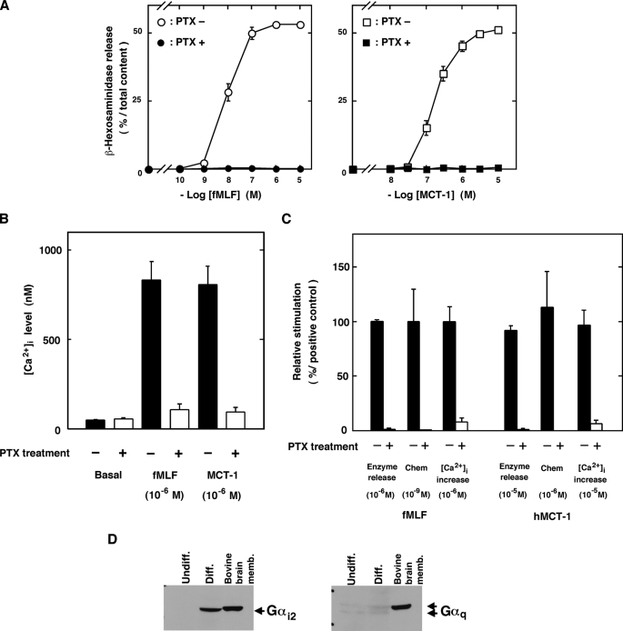
FIGURE 6.
Involvement of Gi protein in MCT-1 signaling. Inhibitory effects of PTX on the stimulation with fMLF, MCT-1, and hMCT-1 (A–C) are shown. A, β-hexosaminidase release from cells treated with 50 ng/ml PTX for 16 h (closed symbols) or from control cells (open symbols) after stimulation with various concentration of fMLF (left, circles) or MCT-1 (right, squares). B, [Ca2+]i increase induced by fMLF or MCT-1 in cells treated with 50 ng/ml PTX for 16 h (open columns) or in control cells (filled columns). C, abilities of hMCT-1 and fMLF to induce β-hexosaminidase release, chemotaxis, and increases in [Ca2+]i were measured in cells treated with 50 ng/ml PTX for 16 h (open columns) or in control cells (filled columns). In C, the effects of fMLF and hMCT-1 on PTX-treated cells are expressed as percentages of those induced by fMLF in the control cells. All data are expressed as mean ± S.E. of five independent experiments. Chem, chemotaxis. D, the expression of Gαi2 (left) or Gαq (right) proteins in the undifferentiated (Undiff.) or differentiated (Diff.) cells was analyzed on Western blots using specific antibodies (see “Experimental Procedures”). As positive controls, Gαi2 and Gαq proteins in bovine brain membrane were also visualized using the same specific antibodies. memb., membrane.

