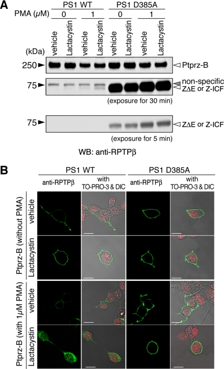
FIGURE 9.
Intramembrane cleavage of Ptprz in HEK cells stably expressing presenilin or its dominant-negative variant. A, HEK293 cells stably expressing either wild-type presenilin 1 (PS1 WT) or a dominant-negative PS1 variant (PS1 D385A) were transiently transfected with the Ptprz-B expression construct. Twenty-four hours after transfection cells were treated and analyzed by Western blotting (WB) using anti-RPTPβ as described in Fig. 8. The figure is representative of three separate experiments, and the result of the densitometric analysis is shown in supplemental Fig. S5. The designations are shown in Fig. 1, A and E. B, the cells treated as described in A were fixed and stained with anti-RPTPβ (green). Merged images with TO-PRO-3-stained nuclei (red) and differential interference contrast images (DIC) are also shown. Scale bars, 10 μm. The figures are representative of three separate experiments.

