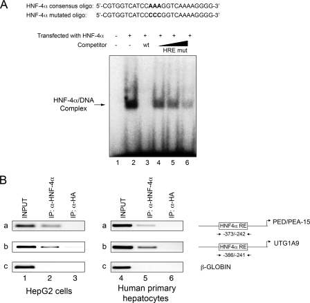
FIGURE 4.
HNF-4α binding to the PED/PEA-15 gene promoter. A, electrophoretic mobility shift assay. Whole cell extracts from HeLa cells transfected with either the empty plasmid (lane 1) or the HNF-4α expression vector (lanes 2-6) were incubated with the 32P-labeled HNF-4α RE probe. Incubation occurred in the absence (lanes 1-2) or the presence of either a 4-fold molar excess of unlabeled HNF-4α RE probe (HRE wt; lane 3) or a 2-, 4-, 10-fold molar excess of unlabeled HNF-4α RE mutated oligonucleotides (HRE mut; lanes 4-6). Proteins were separated on a non-denaturing polyacrylamide gel and revealed by autoradiography. The autoradiograph shown is representative of four independent experiments. B, ChIP assay. Soluble chromatin was prepared from HepG2 cells and human primary hepatocytes as described under “Experimental Procedures” and immunoprecipitated (IP) with either HNF-4α (lanes 2 and 5) or HA antibodies (lanes 3 and 6). Total (INPUT, lanes 1 and 4) and immunoprecipitated DNAs were then amplified using primer pairs covering HNF-4α RE on the PED/PEA-15 (a) and the UGT1A9 promoters (b, positive control) or the β-GLOBIN (c, negative control). The photographs shown are each representative of three independent experiments.

