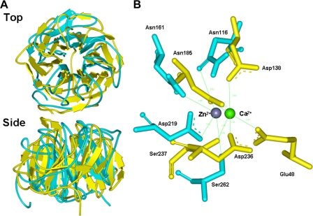
FIGURE 5.
Structural comparison of Staphylococcus Drp35 and predicted model of Euglena ALase. A, superim-position of Staphylococcus Drp35 and Euglena ALase. The protein structure of Drp35 and ALase is shown in yellow-green and pale blue, respectively. B, Ca2+ binding center in Drp35 and prediction of Zn2+-binding residues in Euglena ALase. The residues in Drp35 and ALase are shown in yellow-green and pale blue, respectively.

