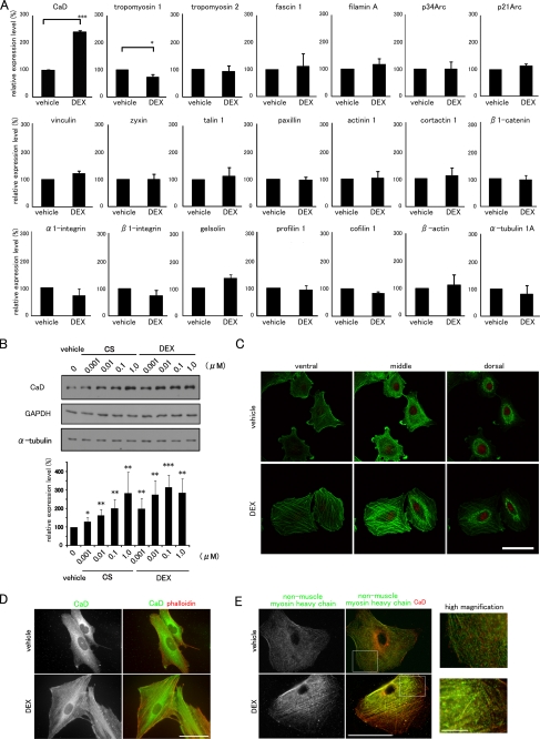
FIGURE 2.
CaD is specifically up-regulated by GCs and localizes along thick stress fibers. A, effects of DEX on the expression profiles of actin cytoskeletal genes. Real time qPCR of cDNAs made from A549 cells. The mRNA expression levels of actin-interacting proteins (CaD, tropomyosin 1, tropomyosin 2, fascin 1, filamin A, p34Arc, p21Arc, vinculin, zyxin, talin 1, paxillin, α-actinin 1, cortactin 1, β1-catenin, α1-integrin, β1-integrin, gelsolin, profilin 1, cofilin 1, β-actin, and tubulin α1a) are shown. The data were obtained from three independent experiments (mean ± S.D., *, p < 0.05; ***, p < 0.001). B, GCs induced the up-regulation of the CaD protein in A549 cells. The cells were incubated with cortisol (CS) or dexamethasone (DEX) at the indicated concentrations for 48 h. The expression levels of CaD, α-tubulin, and GAPDH were determined by Western blot analysis. The CaD protein levels were quantified using ImageJ and normalized to α-tubulin expression. (mean ± S.D., *, p < 0.05; **, p < 0.01; ***, p < 0.001 compared with vehicle). C, confocal images of DEX-induced reorganization of the actin cytoskeleton. Vehicle- or DEX-treated A549 cells were stained with phalloidin (green) and propidium iodide (PI) (red). Bar, 50 μm. D, DEX-induced reorganization of the actin cytoskeleton. Vehicle- or DEX-treated A549 cells were stained with anti-CaD antibody (green in merged image) and phalloidin (red in merged image). Bar, 50 μm. E, DEX-induced stress fiber formation. Vehicle- or DEX-treated A549 cells were pretreated with 0.1% Triton X-100 in PBS and fixed, followed by staining with anti-CaD (red in merged image) and anti-non-muscle myosin heavy chain (green in merged image) antibodies. Bar, 50 μm. Areas within the insets are shown at higher magnification (right panels, Bar, 10 μm).

