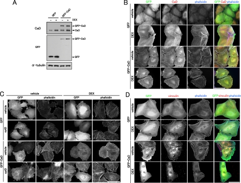
FIGURE 7.
Effect of forced expression of GFP-CaD on the actin cytoskeleton. A, forced expression of GFP-CaD. The expression levels of GFP-CaD, control GFP, and endogenous CaD in A549 transfectants were analyzed by Western blotting with anti-CaD or anti-GFP antibody. B, effect of DEX on the actin cytoskeleton of A549 cells stably expressing GFP or GFP-CaD. The cells were stained with anti-GFP (green in merged image) and anti-CaD (red in merged image) antibodies and phalloidin (blue in merged image). Bar, 50 μm. C, effect of forced expression of GFP-CaD on stress fiber stability to cytochalasin D (cytD). GFP- or GFP-CaD-expressing A549 cells were incubated with vehicle or 1 μm DEX for 48 h. The cells were treated with or without 0.2 μm cytD for 20 min, and stained with anti-GFP antibody and phalloidin. Bar, 50 μm. D, effect of DEX on focal adhesion assembly of A549 cells stably expressing GFP or GFP-CaD. The cells were stained with anti-GFP (green in merged image) and anti-vinculin (red in merged image) antibodies and phalloidin (blue in merged image). Bar, 50 μm.

