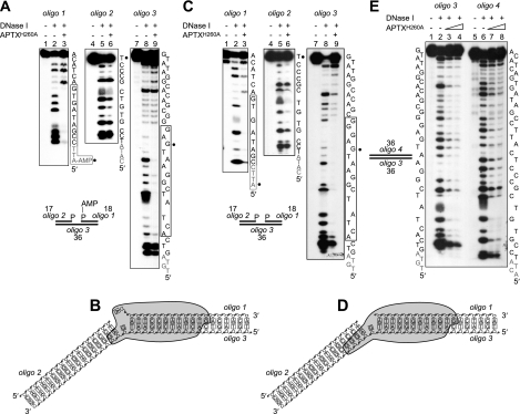
FIGURE 2.
DNase I footprinting of APTX-DNA complexes. A, adenylated SSB DNA 5′-32P-end-labeled on strand 1, 2 or 3 as indicated was incubated with DNase I in the presence or absence of APTXH260A (50 nm) as described under “Experimental Procedures.” Reaction products were resolved by 12% denaturing PAGE and visualized by autoradiography. Nucleotides protected from DNase I are indicated with a box. Gray font denotes 5′-terminal nucleotides not resolved by PAGE; black circles mark the position of the nick. B, graphic representation of the binding of APTX (indicated as a gray shape) to the adenylated nicked DNA. C and D, same as A and B except the DNA was non-adenylated. E, same as A except non-adenylated linear duplex DNA was used. APTX was present at 50 and 100 nm.

