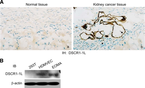
FIGURE 1.
Expression of DSCR1-1L in endothelial cells. A, immunohistochemical staining of a control human kidney tissues (a) and human kidney adenocarcinoma (b) with an antibody specific for human DSCR1-1L. DSCR1-1L expressed in vessels (arrow), not in tumor cells (arrowhead). Tissue from 1 of 5 different patients, all of which exhibited similar staining, is shown. B, cellular extracts from 293T cells, HMDVEC, and EOMA cells were immunoblotted with antibodies against DSCR1-1L (top panel) and β-actin (bottom panel) as a protein equal-loading control.

