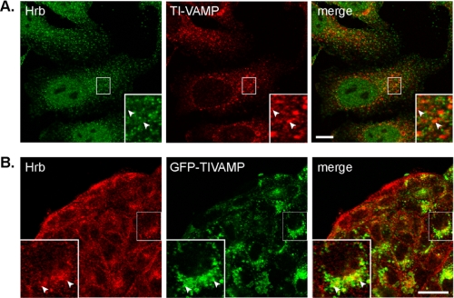
FIGURE 3.
Localization of TI-VAMP and HRB in HeLa and MDCK cells. A, HeLa cells were fixed and stained for endogenous HRB (in green) and TI-VAMP (in red). Arrowheads point to structures positive for only one of the proteins. No significant colocalization was observed at steady state, indicating that HRB and TI-VAMP could interact transiently (bar, 10 μm). B, MDCK cells expressing inducible GFP-TI-VAMP were fixed and stained for the endogenous HRB (in red). Arrowheads point to structures labeled by both HRB and GFP-TI-VAMP (bar, 10 μm).

