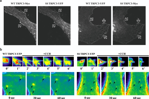
FIGURE 3.
Confocal localization of WT and SS TRPC3. a, confocal microscopy of HeLa cells co-transfected with Myc-tagged full-length WT and YFP-tagged SS TRPC3 or vice versa. Arrows depict areas of co-localization. b, TIRF of HEK293 cells stably transfected with either YFP-tagged full-length WT or SS TRPC3. These cells were stimulated with 100 μm CCH over a time course. Arrows demarcate TRPC3-containing vesicles and their movement over time. See supplemental movies for real-time TIRF experiments.

