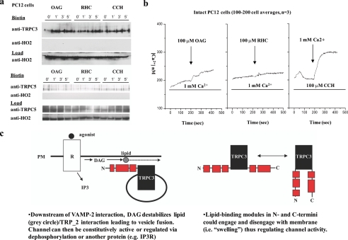
FIGURE 8.
DAG induces fusion of endogenous TRPC3-containing vesicles with the plasma membrane. a, top, Western blot of biotinylated non-transfected rat PC12 cells treated in a time course (0, 1, 3, and 5 min) with 100 μm OAG, 100 μm RHC (a DAG-kinase inhibitor which increases endogenous levels of DAG), or 100 μm CCH and blotted for endogenous TRPC3. Bottom, Western blot as above except blotted for endogenous TRPC5. b, free Ca2+ measurements were made in non-transfected rat PC12 cells. Cells were acclimated in normal 1 mm Ca2+ containing medium and then challenged with either left: 100 μm OAG (arrow) or middle: 100 μm RHC (arrow). Right:Ca2+ pools were released in cells by CCH (100 μm) (first bar), in nominally Ca2+-free medium followed by replacement with CCH and 1 mm Ca2+-containing media (arrow). These traces represent averages of 100-200 cells. c, oversimplified graphical representation of the proposed fusion mechanism of WT TRPC3.

