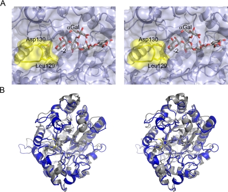
FIGURE 3.
Active center topography of a mannobiohydrolase. A, the solvent-accessible surface of the substrate-binding region of CjMan26C is shown (pale blue) in divergent (wall-eyed) stereo, with the 6163-Gal2Man4 ligand in licorice. Leu-129 and Asp-130, part of the loop which confers exo-specificity, are shaded yellow. B, overlap of the exo-enzyme CjMan26C (gray, the E338A complex with mannobiose for clarity) with the endo-enzyme CjMan26A (blue, with the “Michaelis complex” ligand, DNP-2F mannotrioside, in cyan). The CjMan26C “exo loop” is colored yellow. This figure was drawn with PyMol (DeLano Scientific LLC; available on the World wide Web).

