FIGURE 7.
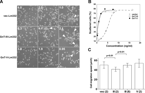
Cell scattering and migration activities of N-glycosylated laminin-332s. BLR cells were incubated with the indicated concentrations of Lm332s. After 40 h of incubation, cells were fixed and stained with crystal violet for observing cell scattering. A, cell morphology of BLR cells in each indicated condition. The numbers on the left are the concentrations for Lm332 (ng/ml) in cell culture medium. The arrowheads indicate the cluster of cells. B, scattered cells were counted and indicated as a percentage against total number of the cells. Crosses, vector-Lm332; open squares, GnT-III-Lm332; closed circles, GnT-V-Lm332. C, effects of vector-Lm332 (vec), GnT-III-Lm332 (III), and GnT-V-Lm332 (V) on migration of Lm332-null keratinocytes. The migration on each substrate was monitored by time lapse microscopy for 8 h. Each bar represents the mean ± S.D. of the migration speeds of nine cells. The numbers in parentheses are the concentrations for Lm332 (μg/ml). Other experimental conditions are under “Experimental Procedures.”

