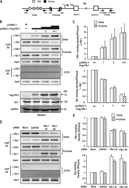
FIGURE 5.
ChIP assays. FBI-1 and Sp1 compete with each other for binding to FRE3 and GC-box 2 in vivo. A, structure of human Rb gene. Arrows, primers used in ChIP; Tsp (+1), transcription start site. Circles with number, FBI-1 binding FREs; filled black circles with number, GC-box that binds Sp1; open boxes, exons; 3′-UTR, 3′-untranslated region with no known binding sites for either Sp1 or FBI-1. B, ChIP assays of binding competition between Sp1 and FBI-1 for the distal (bp –370 to –147), proximal promoter (bp, –131 to +93), and 3′-UTR elements (bp +176326 to +176626) of the endogenous Rb gene promoter in HEK293A cells. Human HEK293A cells were transfected with increasing amount of FLAG-FBI-1 expression vector and analyzed for Sp1 and FBI-1 binding using the antibodies indicated. Sp1 and FBI-1 compete with each other both on the distal and proximal promoter regions. C, histogram of the ChIP assay shown in B. D, ChIP assays on the endogenous Rb gene promoter after knockdown of endogenous FBI-1 expression in HEK293A cells. Knockdown of FBI-1 resulted in an increase in Sp1 binding to the endogenous Rb gene promoter, both on the proximal and distal promoters. E, histogram of the ChIP assay shown in C.

