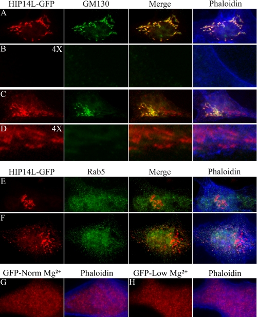
FIGURE 6.
Subcellular redistribution of HIP14L-GFP fusion protein in response to magnesium. Immunofluorescence staining of HIP14L-GFP fusion protein of transiently expressing MDCK epithelial cells. A, Golgi localization of HIP14L-GFP. Cells were cultured in media containing normal magnesium concentration, fixed, and incubated with GFP antibody (HIP14L-GFP) and the Golgi marker, GM130 (GM130). The merged image demonstrates HIP14L-GFP and GM130 colocalization (Merge). A phaloidin overlay of the merged image shows the surface membrane (Phaloidin). B, there was very little submembrane HIP14L protein in MDCK cells cultured in normal magnesium media. The image is of A digitally enlarged 4 times with each of the respective stains. C, Golgi localization of HIP14L-GFP in cells cultured in low magnesium media. Cells were fixed and incubated with GFP antibody (HIP14L-GFP), GM130 (GM130), GFP and GM130 merged (Merge), and phaloidin (Phaloidin). Note, the increase in Golgi HIP14L-GFP and the evident appearance of subplasma membrane HIP14L-GFP protein. D, submembrane location of post-Golgi HIP14L-GFP protein with low magnesium. The image is of C digitally enlarged 4 times with each of the respective stains. Note, the predominant submembrane localization of post-Golgi HIP14L-GFP protein. E, absence of HIP14L-GFP protein in early recycling endosomes supporting the notion that the HIP14L protein does not traffic to the plasma membrane. MDCK cells were cultured in normal media with normal magnesium concentration. The images comprise HIP14L-GFP staining (HIP14L-GFP), Rab5 (Rab5), HIP14L-GFP and Rab5 merged image (Merge), and the phaloidin overlay image (Phaloidin). F, absence of HIP14L-GFP protein in early recycling endosomes. MDCK cells were cultured in media with low magnesium. HIP14L-GFP staining (HIP14L-GFP), Rab5 (indicated as Rab5), merged image (Merge), and the phaloidin overlay image (Phaloidin) are shown. G, control MDCK cells transfected with GFP alone. H, magnesium-restricted MDCK cells transfected with GFP alone. Changes in magnesium concentration do not alter GFP distribution. All images are representative in excess of 30 cells for each condition.

