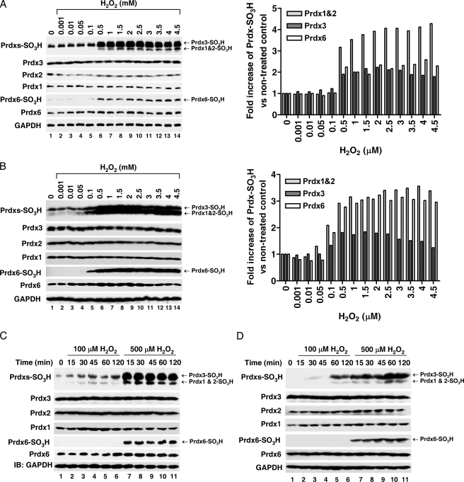
FIGURE 2.
Prdx6 hyperoxidation is dependent on the concentration of H2O2. HeLa (A) and HEK293 (B) cells were treated with different concentrations of H2O2, as indicated, for 20 min. Cell lysates were subjected to immunoblotting with an anti-Prdxs-SO3H antibody that recognizes Prdx1-, Prdx2-, and Prdx3-SO3H, or with anti-Prdx6 SO3H, anti-Prdx1, anit-Prdx2, anti-Prdx3, anti-Prdx6, or anti-GAPDH antibodies. Each graph (left) represents the band intensity quantified by densitometric analysis (ImageJ 1.40 software). HeLa (C) and HEK293 (D) cells were treated with different concentrations of H2O2, as indicated, for different amounts of time. Cell lysates were subjected to immunoblotting as described in A and B.

