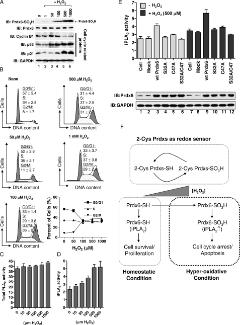
FIGURE 4.
Prdx6 hyperoxidation plays a role in H2O2-induced cellular toxicity. A, HeLa cells were treated with different concentrations of H2O2 as indicated for 20 min, washed with HBSS, and further incubated for 18 h. Cell lysates were subjected to immunoblotting with anti-Prdx6 SO3H, anti-Prdx6, anti-p53, anti-p21, anti-cyclin B1, or anti-GAPDH antibodies. B, after treatment with different concentrations of H2O2 as described in A, cells were washed with HBSS, further incubated for 18 h, stained with propidium iodide as described under “Experimental Procedures”, analyzed with the FACSCalibur™ system, and then cell cycle distributions were determined with the Modfit LT 3.0 software. The results are expressed the mean ± S.D. for triplicate assays. C and D, HeLa cells were treated with different concentrations of H2O2 for 20 min, scraped into Eppendorf tubes, and centrifuged at 1000 × g for 5 min. Total PLA2 (C) and iPLA2 activities (D) were measured as described under “Experimental Procedures.” The results are expressed as mean ± S.D. for triplicate assays. E, HeLa cells were transiently transfected with wild-type Prdx6, the C47A mutant, the C32A mutant, and the C32A/C47A double mutant of Prdx6. At 36 h after transfection, cells were treated with or without 500 μm H2O2 for 20 min. Cell lysates were subjected to immunoblotting with anti-Prdx6 and anti-GAPDH antibodies, and iPLA2 activity was measured. F, model for the role of Prdx6 hyperoxidation in H2O2-induced cellular toxicity.

