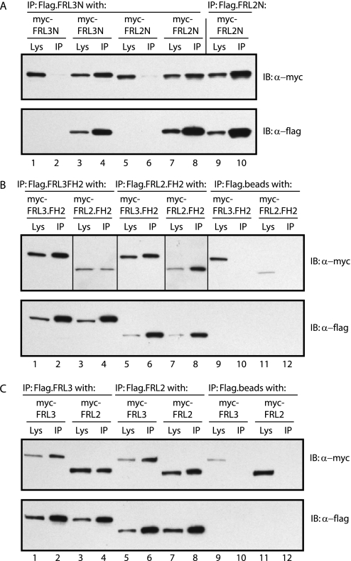
FIGURE 7.
FRL2 and FRL3 are able to form hetero-oligomers. A, FRL2N and FRL3N form hetero-oligomers in vivo. FLAG-tagged FRL3N, or FRL2N, was co-expressed in NIH 3T3 cells with Myc-tagged FRL3N or FRL2N. FLAG-tagged FRL3N efficiently co-immunoprecipitated (IP) FRL3N and FRL2N (compare lanes 1 and 2 with lanes 3 and 4 and lanes 5 and 6 with lanes 7 and 8). FLAG-tagged FRL2N efficiently co-immunoprecipitated FRL2N. FLAG beads alone served as a negative control (lanes 1 and 2 and lanes 5 and 6). B, FRL2.FH2 and FRL3.FH2 form hetero-oligomers in vivo. FLAG-tagged FRL3.FH2 or FRL2.FH2 was co-expressed in NIH 3T3 cells with Myc-tagged FRL2.FH2 and FRL3.FH2. FLAG-tagged FRL3.FH2 efficiently co-immunoprecipitated FRL2.FH2 and FRL3.FH2 (compare lanes 1 and 2 with lanes 3 and 4 and lanes 9 and 10). FLAG-tagged FRL2.FH2 also efficiently co-immunoprecipitated FRL2.FH2 and FRL3.FH2 (compare lanes 5 and 6 with lanes 7 and 8 and lanes 11 and 12). FLAG beads alone served as a negative control (lanes 9-12). Vertical lines separate different exposures of the same immunoblot (lanes 1, 2, 5, 6, and 9-12 versus lanes 3, 4, 7, and 8). C, full-length FRL2 and FRL3 form hetero-oligomers in vivo. FLAG-tagged FRL3 or FRL2 was co-expressed in NIH 3T3 cells with Myc-tagged FRL2 and FRL3. FLAG-tagged FRL3 efficiently co-immunoprecipitated FRL3 and FRL2 (compare lanes 1 and 2 with lanes 3 and 4 and lanes 9 and 10). FLAG-tagged FRL2 also efficiently co-immunoprecipitated FRL2 and FRL3 (compare lanes 5 and 6 with lanes 7 and 8 and lanes 11 and 12). FLAG beads alone served as a negative control (lanes 9-12).

