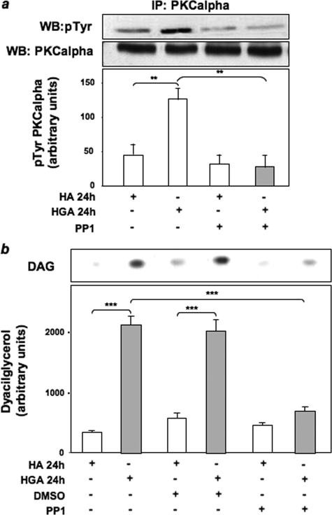
FIGURE 3.
Role of Src in HGA-induced PKCα activation in L6 skeletal muscle cells. a, lysates from L6 skeletal muscle cells treated or not with 0.1 mg/ml HA or HGA for 24 h in the absence or in the presence of 5 μm PP1 were precipitated (IP) with selective anti-PKCα antibodies and immunoblotted (WB) with anti-phosphotyrosine or anti-PKCα antibodies. The autoradiographs shown are representative of three independent experiments. b, L6 skeletal muscle cells were incubated with 0.1 mg/ml HA or HGA for 24 h in the absence or in the presence of 5 μm PP1. Lipids were extracted, and determination of DAG levels was performed as described under “Experimental Procedures.” [32P]Phosphatidic acid was separated by TLC and quantitated by densitometric analysis. Bars represent the mean ± S.D. of values obtained from three independent experiments in duplicate. The autoradiographs shown are representative of three independent experiments. *, statistically significant differences (**, p < 0.01; ***, p < 0.001).

