FIGURE 9.
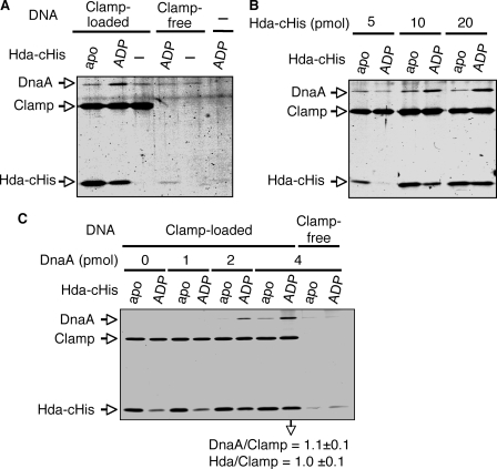
Spin-column analysis for complex formation of ADP-Hda, ATP-DnaA, and the DNA-loaded clamp. Apo-Hda-cHis (apo) and ADP-Hda-cHis (ADP) were prepared as described for Fig. 8A. A, apo-Hda-cHis or ADP-Hda-cHis (10 pmol) was incubated on ice for 5 min in the presence of the DNA-loaded clamp (Clamp-loaded) (1 pmol as clamp, 80 ng as M13mp18 nicked circular DNA) or M13mp18 nicked circular DNA (Clamp-free) (80 ng). ATP-DnaA (2 pmol) was then added to the reaction, followed by immediate spin-column gel filtration, isolation of the void fraction, and analysis by SDS-PAGE described for Fig. 8A. –, no DNA nor protein. B, indicate amounts of apo-Hda-cHis or ADP-Hda-cHis were similarly analyzed using the DNA-loaded clamp (1 pmol as clamp) and ATP-DnaA (2 pmol). C, indicated amounts of ATP-DnaA were similarly analyzed using the DNA-loaded clamp (1 pmol as clamp) and apo-Hda-cHis or ADP-Hda-cHis (20 pmol). Two independent experiments were preformed, and similar data were obtained. Representative data are shown. In the experiments using 4 pmol of ATP-DnaA, the mean molar ratios of recovered DnaA/Clamp (β dimer) and Hda/Clamp were deduced using quantitative standards and are indicated below the gel image.

