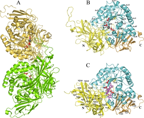
FIGURE 2.
Overall structure of SusB in apo and acrabose complex form. Three catalytic residues, Glu439, Glu508, and Glu532, are shown as stick models in red, and a bound calcium ion is shown as a gray sphere. A, dimer structure of SusB. Mol-A and Mol-B are colored green and khaki, respectively. B, monomer structure of SusB. Domains N, A, and C are shown in yellow, cyan, and gold, respectively (same in C). The secondary structure elements of domain A are labeled following the order of typical (β/α)8 barrel structures, and the prime and double prime refer to the atypical elements. C, monomer structure of SusB with bound acarbose molecule shown as stick model in magenta at the active site pocket. The secondary structure elements of domain N and C are labeled.

