FIGURE 1.
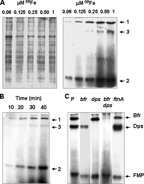
Native PAGE analysis of protein extracts from E. chrysanthemi cells supplied with 59Fe-chrysobactin. Protein-bound iron was detected by autoradiography. Further details are described under “Experimental Procedures.” A, 59Fe-chrysobactin was supplied to parental cells at the indicated concentrations for 40 min. The autoradiogram shown in the right panel corresponds to proteins visualized by Coomassie Blue staining in the left panel. B, 0.25 μm 59Fe-chrysobactin was supplied to parental cells for the indicated times. C, parental and relevant mutant cells (as indicated by P and corresponding genotypes) were supplied with 1 μm 59Fe-chrysobactin for 40 min. Insoluble material is visible at the tops of some lanes. 59Fe-labeled signals are designated by arrows 1, 2, and 3, respectively.

