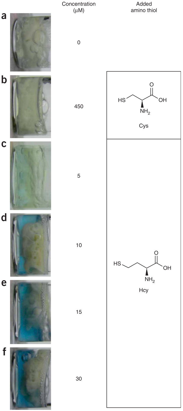
Figure 2.
Photographs of vials containing human blood plasma and methyl viologen dication in buffer solution containing (from upper to lower) (a) no added reduced Hcy, (b) 450 μM cysteine, (c) 5 μM Hcy, (d) 10 μM Hcy, (e) 15 μM Hcy and (f) 30 μM Hcy. The results demonstrate that the color formation and intensity are due to the Hcy levels. Threshold values denoting dangerous Hcy concentrations in human blood plasma are ≥12–15 μM. Normal urinary values might vary but are generally <9.5 μM The shades of the solutions shown are intended as a general guide, along with the protocol, to allow the user to adjust the sensitivity and detection limit. The solid material in the vials apparently comprises denatured proteins and related materials that congeal upon heating.

