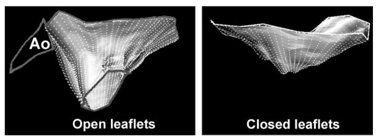
Figure 2.
Three-dimensional reconstructions of total mitral leaflet area in diastole (left) and systolic leaflet closure area (right) in a patient with FMR and both systolic and diastolic leaflet tethering.24a Ao indicates aorta; the left atrium is above, LV below, anterior leaflet to the left.

