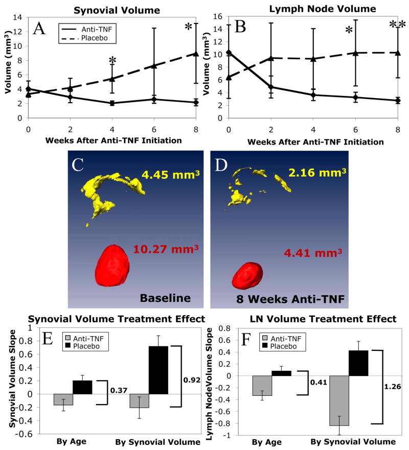
Figure 5. Effects of anti-TNF therapy in TNF-Tg mice with established synovitis.
TNF-Tg mice (n=4) received bimonthly CE-MRI from 3-months of age until they reached a synovial volume above 3mm3. At this time they received an in vivo micro-CT scan and were randomized into anti-TNF and placebo treatment groups (n=4 per group). The mice were then scanned every 2 weeks until sacrifice at 8 weeks, when they received a follow-up micro-CT scan. The synovial (A) and LN (B) volumes for each scan were calculated, and the data from the placebo (dashed line) vs. anti-TNF (solid line) groups, are plotted as the mean +/− standard deviation. Two-sided t-tests revealed significant (*, p<0.05) and highly significant (**, p<0.01) differences between anti-TNF vs. placebo groups at the same time point after therapy. Linear mixed-effects regression analysis revealed a highly significant difference in slopes for anti-TNF versus placebo in both synovial (p<0.001) and LN (p<0.0001) volumes. The protective effects of anti-TNF therapy were also apparent from the Amira 3D reconstructions of pannus and LN, as shown in baseline (C) and 4 weeks (D) images from a representative mouse. Treatment effect was evaluated by a measure of the difference in slopes between anti-TNF and placebo groups. When treatment was initiated using synovial volume rather than age as the entrance criterion, there was a significantly larger treatment effect in synovial volume (E, 0.92 versus 0.37 mm3/week, p=0.04) and in lymph node volume (F, 1.26 versus 0.41 mm3/week, p=0.04).

