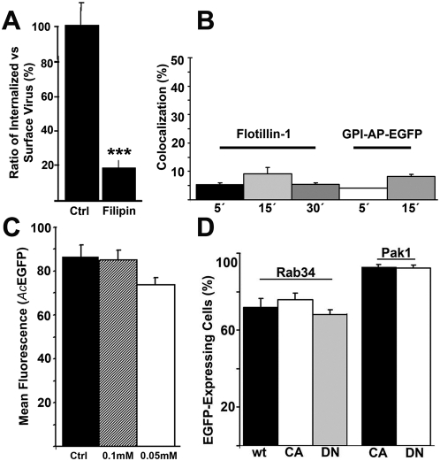
Figure 3. Plasma membrane rafts and macropinocytosis in baculovirus uptake.
(A) The ratio of surface-bound (baculovirus Ab, A555) vs. intracellular baculovirus (baculovirus Ab, A488) was measured after 30 min p.t. during treatment with filipin in HepG2 cells from three separate samples (30 cells). Confocal sections were analyzed by BioimageXD. Mean values and standard deviations are shown. (B) Quantification of baculovirus colocalization with Flotillin-1 or GPI-AP-EGFP were analyzed by confocal microscopy in 293 cells after 5, 15 or 30 min p.t. from 30 cells from three separate experiments. Mean values and standard deviations are shown. (C) Macropinocytosis inhibitor EIPA (0.05 mM and 0.1 mM) was tested for its effects on baculovirus-mediated (Ac-EGFP, MOI 200) EGFP expression. Mean values of fluorescence intensity and standard deviations from FACS analysis are shown. (D) Baculovirus mediated EGFP expression was quantified at 6 h p.t. in the presence of transfected (48 h) macropinocytosis regulators Rab34 and Pak1 in 293 cells. The proportion of nuclei positive for EGFP expression of transfected cells was calculated from two separate experiments by confocal microscopy. In all studies, p-values were determined by unpaired Student's t test with a two-tailed P value. *P<0.05, **P<0.01, ***P<0.001.

