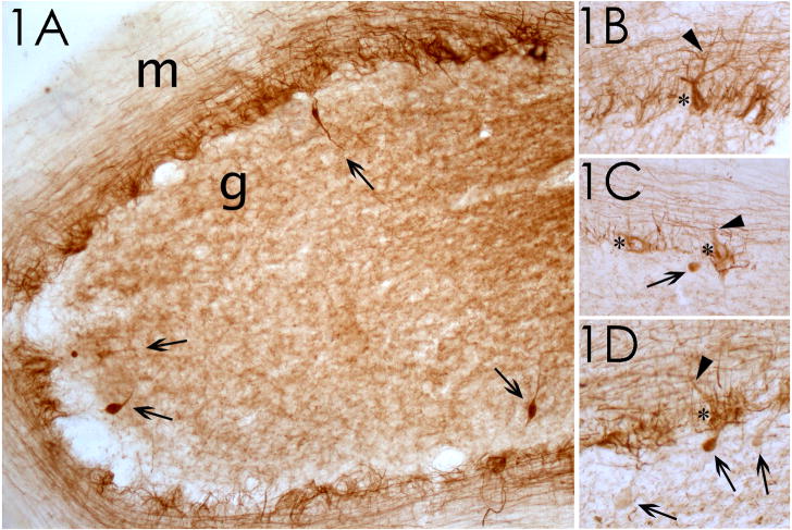
Figure 1.
Phosphorylated neurofilament staining of cerebellum in ET cases. (1A) Case 1; the granular cell layer (g) and molecular layer (m) are identified. 100x magnification. Four torpedoes, in the granular cell layer, are marked by arrows. (1B–1D) Cases 2 and 3; pathologic accumulation of phosphorylated neurofilaments in Purkinje cell body (asterisks) and proximal dendrites (arrowheads). In some instances, the axon of these Purkinje cells contains a torpedo (arrows in 1C, 1D). 200x magnification.

