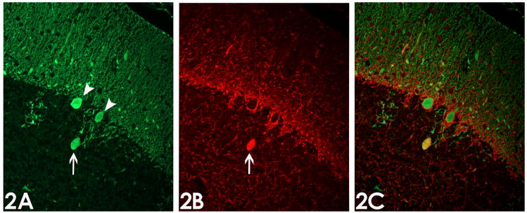
Figure 2.
Immunofluorescence analysis of torpedoes. (2A) Purkinje cell bodies (arrow heads) and torpedo (arrow) are immunoreactive for calbindin. (2B) Torpedo (arrow) but not Purkinje cell bodies are immunoreactive for phosphorylated neurofilament protein. (2C) Overlay of calbindin and phosphorylated neurofilament protein immunostains shows co-localization of these proteins (yellow color). Magnification 200x.

