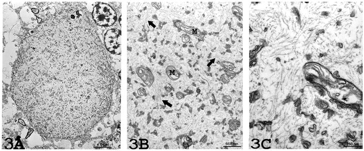
Figure 3.
(3A) Electron microscopy (2,500x magnification) from Case 1 reveals an absence of myelin around the torpedo. The torpedo is comprised of an aggregate of randomly-arranged neurofilaments with scattered organelles. (3B) Higher (15,000x) magnification revealing mitochondria (M) and occasional membrane stacks in a matrix of numerous, disorganized neurofilaments (arrows). (3C) Higher (40,000x) magnification reveals numerous randomly arranged neurofilaments measuring 10 – 12nm nm in diameter.

