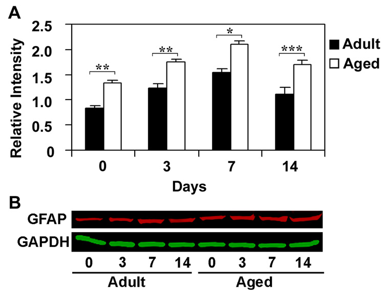
Fig. 6.
Relative expression of GFAP protein in hippocampus of adult and aged mice following CCI injury analyzed using immuoblotting. Protein extracts from hippocampus were analyzed by immunoblotting using anti-GFAP antibodies after 3, 7 and 14 days after CCI injury. Numbers represent relative levels of protein based on optical density normalized to GAPDH signal. The bottom panel shows the representative western blot for GFAP along with GAPDH used as loading control. Data are presented as mean ± SEM (n=3/group). Asterisk denotes statistical significance after ANOVA and post-hoc testing where *p < 0.05, **p < 0.01, ***p < 0.001 vs. adult

