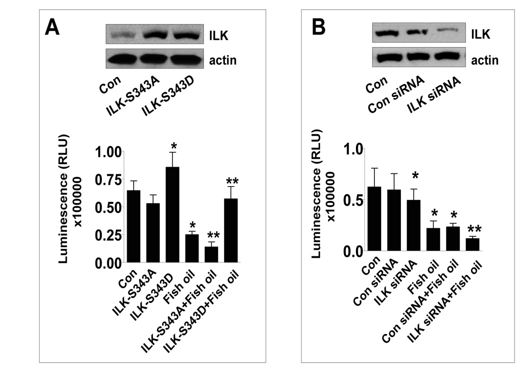
Figure 2. The role of ILK in mediating the inhibitory effect of fish oil on cell growth.

A, H1838 cells were transfected with the inactive (ILK-S343A) and superactive ILK cDNA (ILK-S343D) using the oligofectamine reagent (Invitrogen) according to the manufacturer’s instructions. After 24 h of incubation, cells were treated with or without (15 µg/ml) for an additional 24 h. Afterwards, viable cells were detected using Cell Titer-Glo Luminescent Cell Viability Assay Kit according to the protocol of the manufacturer. The insert in upper panel represents Western blot results for ILK protein. Actin was used as internal control for loading purpose. Con, indicates untreated control cells. B, H1838 cells were transfected with control or ILK siRNA (100 nM) for 40 h followed by exposing the cells to fish oil (15 µg/ml) for an additional 24 h. Afterwards, viable cells were detected using Cell Titer-Glo Luminescent Cell Viability Assay Kit according to the protocol of the manufacturer. The insert in upper panel represents Western blot results for ILK protein. Actin was used as internal control for loading purpose. Con, indicates untreated control cells.
