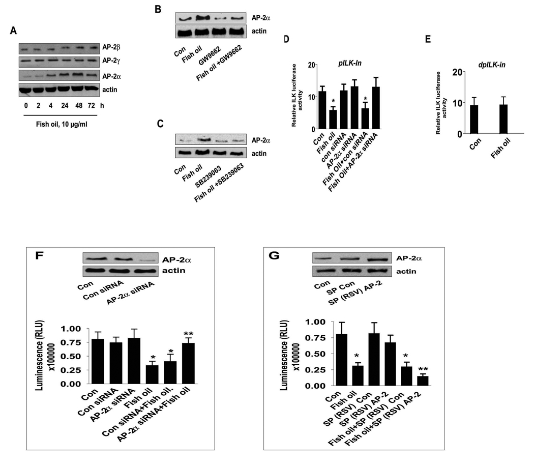
Figure 6. The role of transcription factor AP-2α in inhibition of ILK expression by fish oil.
A, Cellular proteins were isolated from H1838 cells treated with fish oil (10 µg/ml) for the indicated time period. Afterwards, Western Blot analyses were performed using polyclonal antibodies against AP-2α, β, γ. B, H1838 cells were treated with GW9662 (20 µM) for 2 h before exposure of the cells to fish oil (10 µg/ml) for an additional 24 h followed by Western blot analysis for AP-2α protein. C, H1838 cells were treated with SB239023 (10 µM) for 2 h before exposure of the cells to fish oil (10 µg/ml) for an additional 24 h followed by Western blot analysis for AP-2α protein. D, H1838 cells were transfected with control or AP-2α siRNA (100 nM) together with a wild type ILK promoter constructs (−267/+463 bp) for 30 h, followed by exposing the cells to fish oil (10 µg/ml) for an additional 24 h. E, H1838 cells were transfected with a truncated human ILK promoter reporter construct (shown in Fig.4A) ligated to luciferase reporter gene and an internal control phRL-TK Synthetic Renilla Luciferase Reporter Vector as described in Materials and Methods for 24 h using the oligofectamine reagent (Invitrogen) according to the manufacturer’s instructions. After 24 h of incubation, cells were treated with vehicle control (Con) and fish oil (10 µg/ml) for an additional 24 h. Con, indicates untreated control cells. F, H1838 cells were transfected with control or AP-2α siRNA (100 nM) for 30 h before exposure of the cells to fish oil (10 µg/ml) for an additional 48 h. Afterwards, viable cells were detected using Cell Titer-Glo Luminescent Cell Viability Assay Kit according to the protocol of the manufacturer. Actin was used as internal control for loading purpose. Con, indicates untreated control cells. The insert on the top showed the Western blot result for AP-2α protein production. G, H1838 cells were transfected with control and AP-2 expression reporter constructs [SP(RSV)AP-2], and an internal control phRL-TK Synthetic Renilla Luciferase Reporter Vector as described in Material and Methods section for 24 h before exposure of the cells to fish oil (10 µg/ml) for an additional 48 h. The insert in upper panel represents Western blot results for AP-2α protein. Con, indicates untreated cells. The ratio of firefly luciferase to renilla luciferase activity was quantified as described in Materials and Methods. The bars represent the mean ± SD of at least four independent experiments for each condition. * indicates significant increase of activity as compared to controls. ** indicates significance of combination treatment as compared to the nicotine alone (p<0.05). Con, indicates untreated control cells.

