
Figure 2.
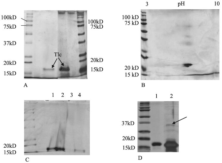
SDS-PAGE of purified Tlc sample separated after ion exchange chromatography and size exclusion chromatography. (A) Comparison of Coomassie blue stained gel (left) with Silver stained gel (right). (B) Silver stained 2D electrophoresis gel of purified Tlc. (C) Coomassie stained gel of Tlc (lane 1) lipidated Tlc (lane 2) delipidated Tlc (lane 3) and relipidated Tlc (lane 4). (D) Delipidated Tlc. A large quantity of delipidated Tlc was applied with extra glycerol in the loading buffer. Lane 1 is Coomassie blue stained and lane 2 silver stained. Arrow indicates a possible dimeric form of the protein.