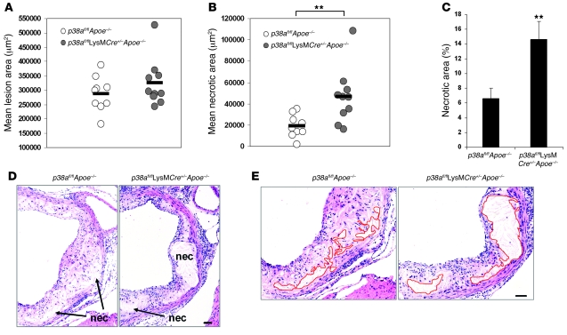
Figure 2. Deficiency in macrophage p38α MAPK increases lesional necrosis.
(A and B) Dot plot showing the aortic root lesion area (A) and the aortic root necrotic area (B) of individual p38afl/flApoe–/– (n = 9) and p38afl/flLysMCre+/–Apoe–/– mice (n = 10). Bars represent mean values. The difference in aortic root lesion area between groups was not significant (P = 0.462, Mann-Whitney U test). (C) Mean percent necrotic area relative to total lesion area for each group. (D) Representative images of aortic root cross sections from p38afl/flApoe–/– and p38afl/flLysMCre+/–Apoe–/– mice stained with H&E. Necrotic areas (nec) are indicated. (E) Images from D, enlarged to demonstrate how the necrotic area was defined for quantification in each section (p38afl/flApoe–/–, 19,018 μm2; p38afl/flLysMCre+/–Apoe–/–, 45,826 μm2). Red lines show the boundary of the developing necrotic core. **P < 0.01, Mann-Whitney U test. Scale bars: 50 μm.

